Technologies
Technical Information
A Bruker BioSpec 94/20 USR 9.4 Tesla multi-purpose MR system equipped with 1H and X-nuclei (carbon, fluorine and phosphorus) radiofrequency coils for different applications is installed at the GMC. A cryogenic radiofrequency coil is also available, especially for brain-imaging applications.
Applications
Obesity and Diabetes Research
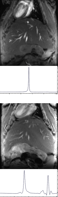
Liver 1H MRS
In the context of diabetes MR methods are applied to study morphological and metabolic changes in different organs. For instance, MRS techniques are used to investigate the deposition of specific fatty acids in the mouse liver during high-fat diet. Changes in the water-fat composition of the liver can be studied by analysing the 1H MRS resonance spectra.
Example of 1H liver spectrum initially acquired at the start of the dietary intervention, showing a single water resonance line at 4.7 ppm
Six months later the increased hepatic lipid content is clearly visible, an additional resonance peak at 1.3 ppm is evident, originating from the methylene (-CH2-)n group
WAT 1H MRS
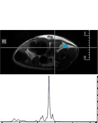
Dietary effects on the fatty acid composition of the adipose tissue (WAT) can be studied with magnetic resonance spectroscopy (1H MRS). An example of a 1H spectra acquired from a volume of interest located in the adipose tissue shows distinct triacylglycerols.
Axial MRI slice through the abdominal region showing the volume of interest (blue square) in which the 1H MRS was performed. Corresponding localized in-vivo 1H spectrum of the adipose tissue, showing the characteristic of a spectrum of triacylglycerol, a large peak at 1.3 ppm originating from methylene (-CH2-)n group .
iBAT 31P MRS
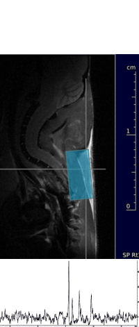
The in-vivo assessment of adenosine-triphosphate (ATP) can be accomplished by spectroscopic approaches. An example of a 31P spectra acquired from a volume of interest located in the interscapular brown adipose tissue (iBAT) is given in the following figure.
Sagittal MRI slice through the neck area showing the volume of interest (blue square, overlaid on the iBAT) . Spectrum of the iBAT showing a large PCr peak, and the γ- α- and β-ATP resonance lines.
Cardiac Imaging
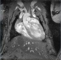
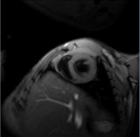
In-vivo cardiac MRI is either performed by synchronizing the data acquisition with the cardiac and respiratory cycle or by using a retrospective gating method (IntraGate™). MR images are acquired at different heart cycles where image planes are generally orientated along the long and the short heart axis.
Cardiac MRI in long axis
Long axis view depicting the four chambers and the major cardiac vessels.
Cardiac MRI in short axis
Short axis view depicting the the left atrium and the left ventricle; images were acquired with dark-blood preparation.
Renal MRI
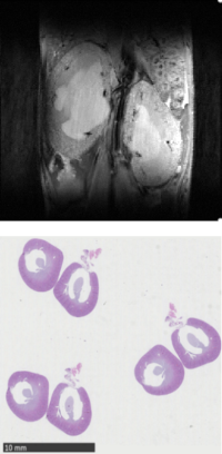
Renal MRI for the analysis of fibrotic and cystic kidney phenotypes.
Example of a kidney phenotype showing severe hydronephrosis (bright signal intensity of pelvis region).
Histopathology. Representative images of HE-stained sections from the affected mutant murine kidney model.
Brain Imaging
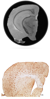
Brain imaging for structural/ functional analysis of neurological phenotypes and pathologies.
Ex-vivo micro-MRI. Example of a murine brain sample revealing decreased signal loss in Aβ plaques due to highly compact fibrilla beta-amyloid masses and/or accumulation of iron, at 31 µm³ spatial resolution.
Histopathology. Representative image of a Congo red/Iba1-stained section from an APP/PS1 mutant murine Alzheimer model (courtesy of K. Niedermeier).